Echocardiography (Echo)
Echocardiography is imaging of the heart with ultrasound (high-frequency sound waves). It is commonly known as an echo. It involves no X-rays, and the structure and movement of the heart are watched live on a screen.
What is assessed?
Echocardiography is one of the most widely used imaging methods in cardiology. It may be requested for shortness of breath, chest pain, palpitations, fainting or a murmur heard during an examination.
- The size of the heart chambers and the thickness of their walls
- The pumping strength of the heart muscle
- The structure of the valves and whether there is narrowing or leakage (regurgitation)
- Fluid build-up around the heart
- Congenital heart disease
- The direction and speed of blood flow inside the heart
Echocardiography through the chest wall
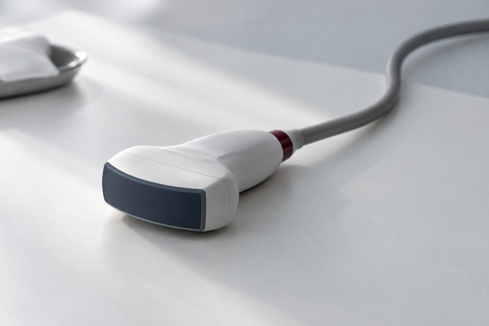
This is the most common type and is called transthoracic echocardiography. You lie on your left side, gel is applied to your chest and a handheld device called a probe is moved over the chest wall. Blood flow is measured in the same test using a technique called Doppler.
The test is painless, you do not need to fast and no special preparation is required. It is usually completed in a short time. You can return to your daily life straight afterwards.
Other types of echocardiography
Other methods may be used when the images taken through the chest wall are not sufficient or when a different question needs answering.
- Echocardiography through the food pipe (transoesophageal): A thin tube with a probe at its tip is passed into the food pipe. It gives more detailed images from close to the heart. You need to fast, your throat is numbed and a sedative is usually given.
- Stress echocardiography: The heart is examined while working under a load created by exercise or medication. It is helpful in investigating coronary artery disease and in some valve conditions.
Interpreting the results
An echocardiography report contains many measurements. One of them, the ejection fraction, shows how much of the blood inside the heart is pumped out with each beat and is a measure of pumping strength.
Some findings, such as a mild valve leak, can also be seen in healthy people and may not mean disease on their own. The results are interpreted by the doctor together with your symptoms, examination and other tests. How often the test is repeated also depends on the underlying condition.
This page is provided for general information. Decisions about diagnosis and treatment are made by a physician after examining you.
Frequently asked questions
Is echocardiography harmful?
Echocardiography through the chest wall uses no radiation and has no known harmful effects. It can also be done in pregnant women and children. There is no objection to repeating it when needed.
Are an echo and an ECG the same thing?
No. An ECG records the electrical activity of the heart and gives information about its rhythm. An echo shows the structure and movement of the heart. The two tests complement each other.
Does echocardiography show blocked arteries?
Echocardiography does not show the inside of the coronary arteries directly. However, it can reveal abnormal movement of the heart muscle caused by artery disease. Other tests are requested when the arteries need to be assessed.
Other areas of expertise
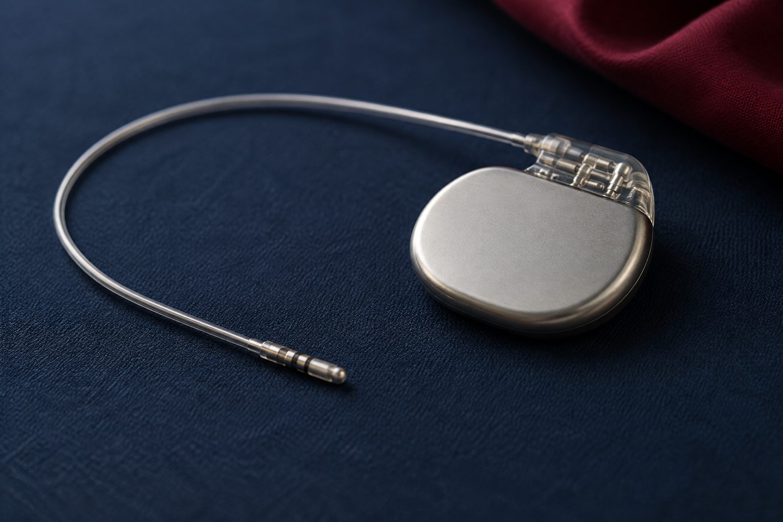 Pacemakers and ICDsDevices placed under the skin that monitor the heart rhythm and deliver electrical impulses when needed.
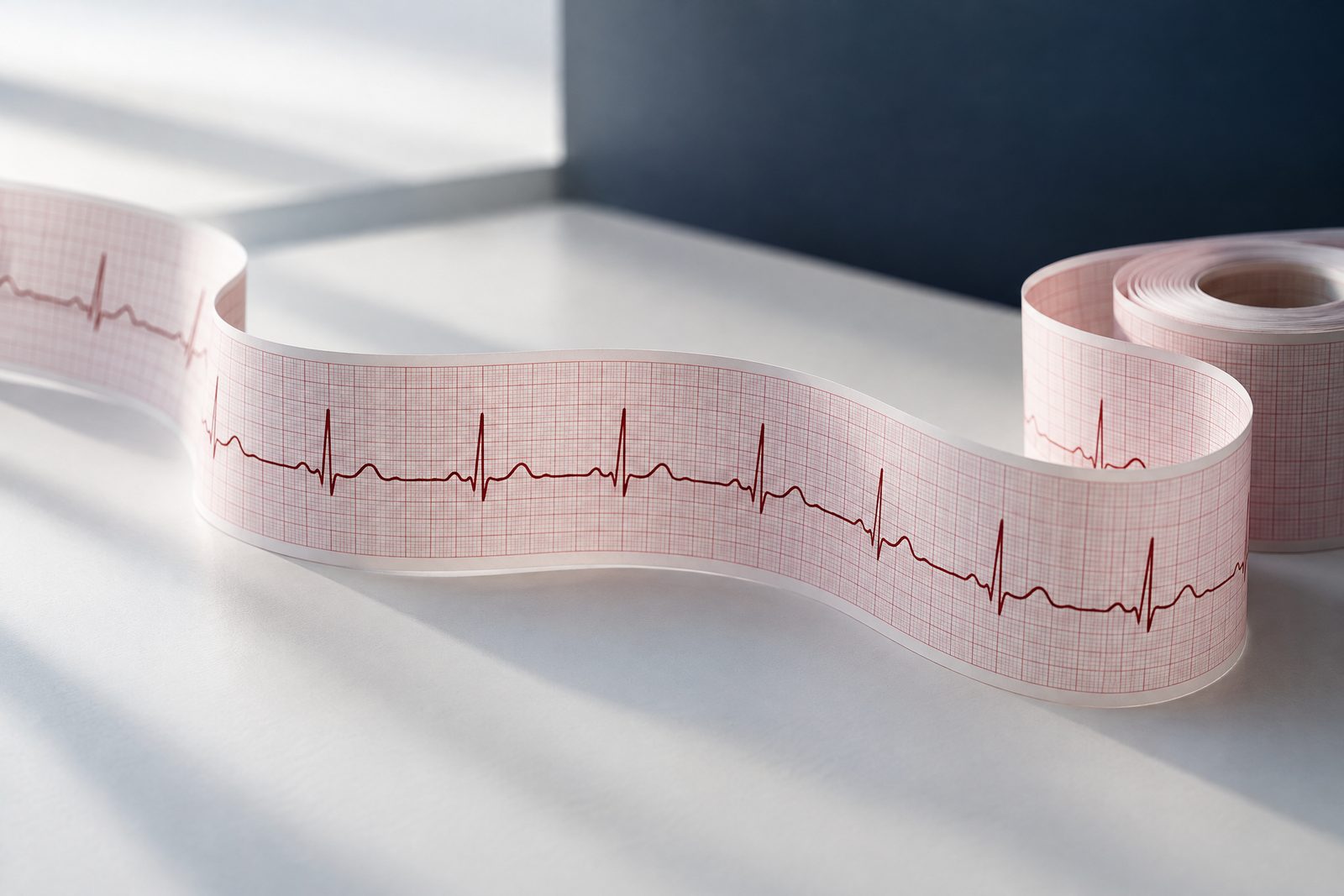 Heart Rhythm DisordersThe general name for conditions that make the heart beat faster, slower or more irregularly than normal.
 HypertensionBlood pressure that stays persistently high. It often progresses without causing symptoms.
Read more on the topic
Short patient information articles on chest pain, blood pressure, diet and cardiac tests.